Comprehensive Eye Care
Services & Procedures
Every service delivered at Neura-Sight is grounded in clinical evidence, modern instrumentation, and a deep understanding of the individual patient in front of us.
Digital Eye Examination
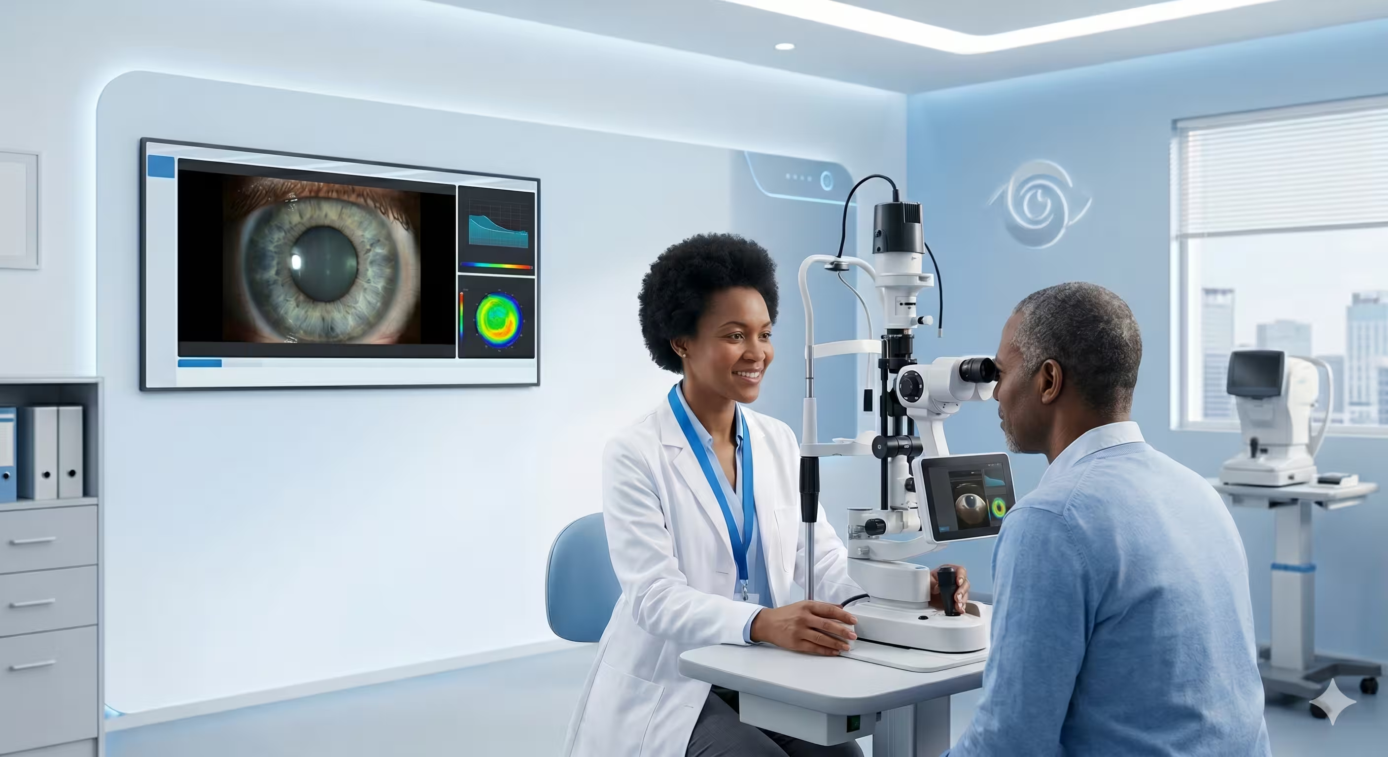
Full anterior and posterior segment evaluation using calibrated digital instruments. The definitive first step in all ocular care pathways.
Refraction & Spectacles
Computerised and subjective refraction, followed by expert dispensing of single-vision, progressive, contact, and driving lenses.
Advanced Procedures
Tonometry for intraocular pressure, fundoscopy for retinal health, and detailed optic nerve assessment with referral pathways when required.
Digital Eye Examination
Our digital eye examination goes far beyond checking whether you need glasses. It is a systematic, evidence-based investigation of the entire visual system — from the surface of the cornea to the furthest reaches of your peripheral retina.
What the Examination Includes
- Case history and visual goals assessment
- Automated refraction using a digital auto-refractometer
- Visual acuity at distance and near (LogMAR chart)
- Colour vision and contrast sensitivity evaluation
- Slit-lamp biomicroscopy: cornea, lens, anterior chamber
- Binocular vision, ocular motility and pupil reflex assessment
- Non-contact tonometry (intraocular pressure screening)
- Posterior segment examination — optic disc, macula, retina
- Clinical findings report and management discussion
Beyond refraction, our clinicians actively screen for early indicators of glaucoma, diabetic changes in the retinal blood vessels, lens opacities, corneal pathology, and any asymmetry that may warrant further investigation.
Book Digital Eye ExaminationRefraction & Spectacle Dispensing
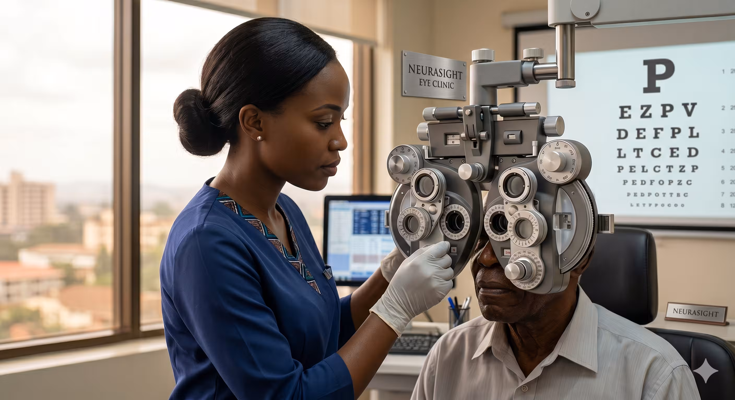
Precision prescription measurement combined with expert lens selection and fitting — every detail of your optical solution is engineered and verified before you leave.
The Refraction Process Explained
Refraction is the clinical procedure used to determine the optical correction your eyes require. At Neura-Sight, we perform both objective refraction (using the auto-refractometer) and subjective refraction (a meticulous clinical process using a digital phoropter and standardised test charts).
The result is a precise, individualised prescription specifying sphere, cylinder, axis, and addition power. We never rely on automated readings alone.
- Objective auto-refraction baseline
- Binocular balance verification
- Stereoscopic depth perception check
- Task-specific near addition refinement for presbyopes
Our Lens Solutions
Single Vision Lenses
Designed to correct a single focal point — distance or near vision. The most commonly dispensed lenses for myopia, hyperopia, and astigmatism.
Materials available: CR-39 organic, polycarbonate, high-index 1.6/1.67, and photochromic variants.
Progressive Addition Lenses (PAL)
A seamless gradient of power providing clear vision at all three distances within a single lens. No visible dividing line.
Our recommendation: We fit premium progressive designs with wide corridors and minimal peripheral swim for first-time wearers.
Contact Lenses
Full fitting service including daily disposable, monthly, toric (astigmatism), multifocal, and specialist scleral lenses for irregular corneas.
Fitting includes: Corneal topography, tear film assessment, insertion training, and 2-week follow-up.
Driving & Specialist Lenses
Polarised anti-glare driving lenses, blue-light filtering for screen users, UV-blocking for outdoor workers, and safety-rated lenses for industrial environments.
Night driving: Anti-reflection coatings on all driving lenses to reduce halo and starburst from headlights.
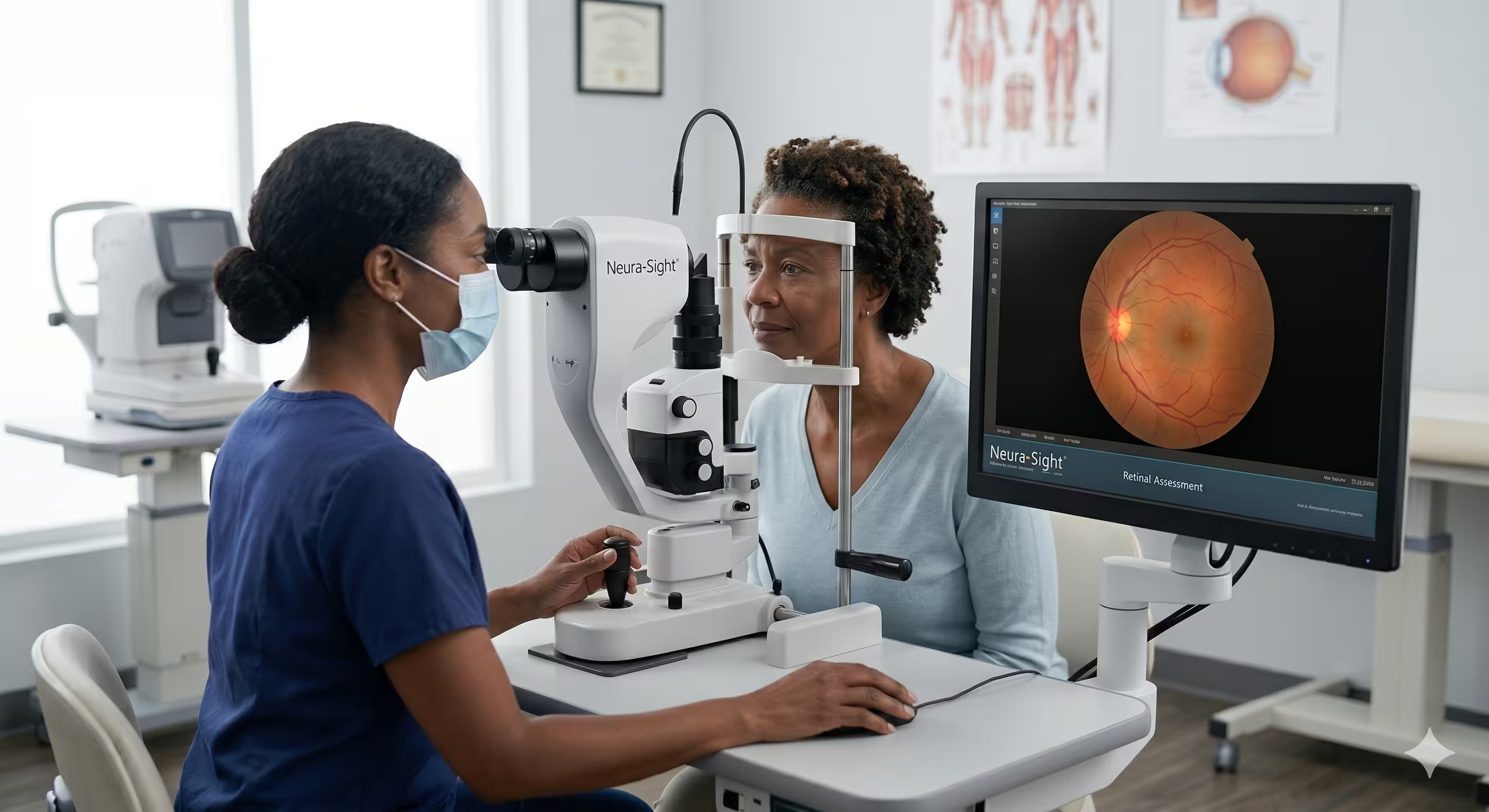
Tonometry & Fundoscopy
Advanced ocular health assessment goes beyond refraction. Our specialists are trained to examine the deep structures of the eye, identifying systemic and ocular disease markers that can be sight-threatening — or life-threatening — if detected late.
Tonometry — Intraocular Pressure Screening
Tonometry is the clinical measurement of intraocular pressure (IOP). Elevated IOP is the primary modifiable risk factor for glaucoma. We use non-contact tonometry (NCT) — a rapid, painless measurement where a brief pulse of air is directed at the corneal surface. No anaesthetic is required and there is no contact with the eye.
- Painless, no contact with the eye surface
- Results available immediately
- Essential for all adults over 40 and anyone with a family history of glaucoma
- Included in all comprehensive eye examinations at Neura-Sight
Understanding your results: Normal IOP ranges from 10–21 mmHg. Readings above this range prompt further investigation including optic nerve head assessment and visual field testing.
Fundoscopy — Retinal Examination
Fundoscopy is the examination of the fundus — the interior surface of the eye, including the retina, retinal vasculature, optic disc, macula, and fovea — using our digital fundus camera and direct ophthalmoscope.
What We Assess During Fundoscopy
- Optic disc: Cup-to-disc ratio, disc margin clarity, and pallor
- Retinal vasculature: AV nicking, haemorrhages, cotton wool spots
- Macula and fovea: Drusen, pigmentary changes, macular oedema
- Peripheral retina: Lattice degeneration, holes, and tears
- Fundus photography: Digital images captured for baseline documentation
Conditions We Diagnose & Manage
Our clinical scope covers the full spectrum of common and complex ocular conditions, from simple refractive errors to sight-threatening retinal disease.
Glaucoma
A group of diseases characterised by progressive optic nerve damage, most commonly associated with elevated intraocular pressure. Entirely symptom-free in its earliest stages — routine pressure checks and disc assessment are the only reliable detection method.
How we screen: Non-contact tonometry, optic disc photography, and visual field analysis.
Diabetic Retinopathy
A complication of diabetes in which elevated blood sugar damages the microscopic blood vessels of the retina, causing progressive vision loss if undetected.
How we screen: Dilated fundoscopy, retinal photography, and referral for OCT imaging when indicated.
Cataracts
Progressive clouding of the crystalline lens causing blurring, glare, and halos around lights. Very common over 60. Modern surgery replaces the cloudy lens with a clear intraocular implant (IOL).
How we screen: Slit-lamp biomicroscopy and visual acuity assessment.
Age-Related Macular Degeneration
A condition affecting the macula — the central, highest-resolution area of the retina responsible for reading and fine detail vision. The leading cause of central vision loss in adults over 55.
How we screen: Fundus photography, Amsler grid testing, and referral for OCT when indicated.
Dry Eye Syndrome
A chronic condition caused by insufficient tear production or poor-quality tear film, causing discomfort, burning, and intermittent blurred vision. Worsened by screen use, air conditioning, and contact lens wear.
How we manage: Meibomian gland assessment, tear film stability testing, and a personalised management protocol.
Keratoconus
A progressive corneal condition in which the cornea thins and bulges forward into a conical shape, causing increasing irregular astigmatism and visual distortion. Onset is typically in the teens and early twenties.
How we screen: Corneal topography, keratometry, and slit-lamp assessment.
Ready for a Full Clinical Evaluation?
Book a comprehensive examination with our specialist team. Complete assessments available Monday through Saturday.